Categories
12th botany neet school

Mutation and classification of mutation

Mutation

Mutation and classification of mutations

Back to botany topic list

In a species, variations are caused by changes in the environment or any changes in the innate genetic setup of an organism or by the combination of both.

Sudden change in the genetical set up of an organism is defined as mutation.

In 1901, Hugo de Vries first used the term mutation based on his observation on Oenothera lamarckiana.

Charles Darwin termed these sudden change as ‘sports’. According to Bateson, mutation is a discontinuous change.

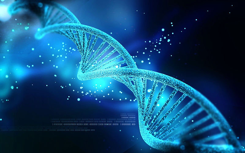
Based on molecular basis of heredity, mutations is defined as sudden change in the sequence of nucleotides of gene.

The mutation brings about a change in the organism. The organism which undergoes mutation, is called a mutant.

eg. Oenothera lamarckiana.

Mutations that affect the biochemical reactions are called biochemical mutations.

For example, biochemical mutants of Neurospora failed to synthesize certain amino acids.

Some mutations drastically influence the genes and cause death to the individual.

Such mutations are described as lethal mutation.

For example, in the plant Sorghum, recessive mutant fails to produce chlorophyll and therefore they die in the seedling stage.

Thus, most of the mutations are harmful, because they disturb the genic balance of the organism.

Although most of the mutations are useless and even harmful, and some of the mutations play a significant role in the evolution of new species.

Many new strains of cultivated crops and new breeds of domesticated animals are the products of gene mutations.

Small seeded Cicer arietinum (bengal gram) suddenly get mutated to large seeded Cicer gigas is the case of gene mutations.

Classification of mutation


Mutations have been classified in various ways based on different criteria.

Depending on the kind of cell in which mutations occur, they are classified into somatic and germinal mutations.

They may be autosomal or sex chromosomal according to their type of chromosome in which they occur.

They may be spontaneous or induced according to their mode of origin.

They may be forward or backward according to their direction.

They may be dominant or recessive according to their phenotypic expression of mutated genes.

Point or gene mutation

Point mutations is sudden change in small segment of DNA either a single nucleotide or a nucleotide pair.

Gross mutations is a change involving more than one or a few nucleotides of a DNA.

The gene mutation may be caused by loss or deletion of a nucleotide pair.

This is called deletion mutations and reported in some bacteriophages.

Addition of one or more nucleotides into a gene results in addition mutations.

Replacement of certain nitrogen bases by another base in the structure of DNA results in substitution mutations.

The deletion and addition mutation alter the nucleotide sequence of genes and ultimately result in the production of defective protein and this leads to the death of the organism.

The substitution mutations can alter the phenotype of the organism and have great genetic significance.

There are two types of substitution mutations – transition and transversion.

When a purine or a pyrimidine is replaced by another purine or pyrimidine respectively this kind of substitution is called transition.

When a mutation involves the replacement of a purine for pyrimidine or viceversa this is called transversion.

Back to botany topic list

For more details about about mutations click here

Other links 

STRUCTURE OF CHROMOSOME – CELL BIOLOGY


Types of chromosomes with special types


Gene and genome


Linkage and mechanism of linkage


Crossing over, gene mapping and recombination of chromosome


Mutagenic agents and its significance


Structural Chromosomal aberrations


Numerical chromosomal aberrations


Role of DNA 


Structure of DNA and Function of DNA


Replication of DNA


Structure of RNA and Types of RNA

Categories
12th botany neet school

Crossing over, gene mapping and recombination of chromosome

Crossing over, gene mapping and recombination of chromosome

Crossing over, gene mapping and recombination of chromosome are explained in detail.

Crossing over

Back to botany topic list

The process, which produces recombination of genes by interchanging the corresponding segments between nonsister chromatids of homologous chromosomes, is called crossing over.

A crossing over between linked genes allows their recombination during meiosis.
Crossing over takes place in pachytene stage of prophase I of meiosis.

In pachytene stage, the bivalent chromosome becomes tetrad i.e. with four chromatids.

The adjacent nonsister chromatids are joined together at certain points called chiasmata.

Crossing over occurs between the nonsister chromatids of paired chromosomes in the region of chiasma.

At each chiasma, the two nonsister chromatids break, exchange their segments and rejoin resulting the crossing over.

crossing over
crossing over

Hence, out of four chromatids the two adjacent chromatids are recombinants and other two are original chromatids.

Thus four types of gametes are obtained.

Significance of crossing over

Crossing over leads to the production of new combination of genes and provides basis for obtaining new varieties of plants.

It plays an important role in the process of evolution.

The crossing over frequency help n the construction of genetic maps of the chromosomes.

It gives us the evidence for linear arrangement of linked genes in a chromosome.

Gene mapping

Genes are arranged linearly in a chromosome. The point in a  chromosome where the gene is located is called locus.

The diagrammatic representation of location and arrangement of genes and relative distance between linked genes of a chromosome is called linkage or genetic map.

The unit of genetic map is Morgan or centimorgan.

When the percentage of crossing over between two linked genes is 1 per cent, then the map distance between the linked genes is one morgan.

There is a greater probability of occurrence of crossing over, when the two genes are farther apart in a chromatid.

The probability of crossing over between two genes is directly proportional to the distance between them.

When two genes are nearer, the probability of occurrence of crossing over between them is limited.

Let A, B, C, D and E be five knots on a string separated by the distances as shown.

The probability of making a random cut between two knots is directly proportional to the distance between them.

Every cut separates A from E, whereas 5/100t1, cut only separates C from D.

If the knots or genes linearly arranged on a chromosome in randoms are the cross overs, then C and D remain linked, whereas A and E will not show linkage in this situation.

Uses of gene mapping

It is useful to determine the location, arrangement and linkage of genes in a chromosome.

It is useful to predict the results of dihybrid and trihybrid crosses.

Recombination of chromosome

The process, which produces recombinations of gene by interchanging of corresponding segments between nonsister chromatids of homologous chromosomes, is called recombination of chromosomes.

It takes place in pachytene stage of prophase I of meiosis. Crossing between the linked genes results in genetic recombination.

According to Bateson and Punnet, in Lathyrus odoratus 12 per cent of the test cross progeny were recombinants.

Recombination between two genes is expressed in percentage. It is called recombination frequency.

Gene pairs that had very low percentage of recombination are known as tightly linked genes.

The gene pairs with higher percentage are termed as loosely linked genes.

For example, 12 per cent of the test cross progeny were recombinants.

They showed a different linkage of alleles than their parents.

The percentage recombination is determined by dividing the number of recombinant offspring by the total number of offspring.

In the figure 3.4, the linkages of the parents were B with L and b with l.

The recombinant offspring are B with l or b with L.

Back to botany topic list

For more detail about cross over click here

Other links 

STRUCTURE OF CHROMOSOME – CELL BIOLOGY


Types of chromosomes with special types


Gene and genome


Linkage and mechanism of linkage


Mutation and classification of mutation


Mutagenic agents and its significance


Structural Chromosomal aberrations


Numerical chromosomal aberrations


Role of DNA 


Structure of DNA and Function of DNA


Replication of DNA


Structure of RNA and Types of RNA

Categories
12th botany neet school

Linkage and mechanism of linkage

Linkage and mechanism of linkage

Linkage and mechanism of linkage

Back to botany topic list

The tendency of genes or characters to be inherited together because of their location on the same chromosome is called linkage.

Many hybridization experiments were conducted both on plants and animals based on Mendel’s work.

The results of certain dihybrid crosses did not confirm the law of independent assortment.

It states that the inheritance of genes of each pair in a dihybrid during gamete formation is independent of the other.

linkage - mechanism of linkage

In 1906, William Bateson and Reginald Punnett conducted experiments in sweet pea, Lathyrus odoratus to confirm Mendel’s dihybrid testcross.

They observed an exception to the independent assortment of two genes in this plant.

Here, blue flower (B) is dominant over the red flower (b) and long pollen (L) dominant over round pollen (l).

They crossed true breeding plants having blue flower with long pollen (BBLL) and red flower with round pollen (bbll). All the F1 hybrids have blue flowers with long pollen (BbLl).

A testcross between heterozygous blue long (BbLl) of Fl hybrid and double recessive parental stock red round (bbll) did not result in ratio 1:1:1:1 but gave unexpected phenotype frequency as shown below.

Here, blue long and red round are parental forms and show greater frequency 88 per cent.

Blue round and red long are recombinant forms and show lesser frequency 12 per cent. The dihybrid test cross ratio obtained is 7:1:1:7 and not 1:1:1:1.

This indicates that the genes do not independently assort. From the above test cross, it is clear that if dominant alleles or recessive alleles are present in the same plant, they tend to remain together resulting in increased parental forms.

Thus, the two genes which inherit together are called linked genes. This aspect is called coupling.

They made another cross between plants having blue flower with round pollen (BBll) and red flower with long pollen (bbLL). A testcross between
Mechanism of linkage

heterozygous blue long (BbLl) of Fl hybrid and double recessive red round (bbll) did not result in ratio 1:1:1:1 but gave unexpected phenotype frequency as shown below.

Here, blue round and red long are parental forms and show greater frequency 88 per cent.

Blue long and red round are recombinant forms and show lesser frequency 12 per cent.

The dihybrid test cross ratio obtained is 1:7:7:1 and not 1:1:1:1. This indicates that the genes do not independently assort.

From the above testcross, it is clear that if dominant alleles or recessive alleles are present in the different plants, they tend to remain separate resulting in increased parental forms.

This aspect is called repulsion.

Coupling and repulsion offered explanation for higher frequency of parental forms.

They are two aspects of a single phenomenon called linkage. The genes that are carried on the same chromosome will not assort independently because of their tendency to remain linked together.

This is called linkage. The genes located on the same chromosomes that are inherited together are known as linked genes.

They tried to reconfirm the law of independent assortment.

But they could not get expected result because the genes are linked

Back to botany topic list

For more details about linkage click here

Other links 

STRUCTURE OF CHROMOSOME – CELL BIOLOGY


Types of chromosomes with special types


Gene and genome


Crossing over, gene mapping and recombination of chromosome


Mutation and classification of mutation


Mutagenic agents and its significance


Structural Chromosomal aberrations


Numerical chromosomal aberrations


Role of DNA 


Structure of DNA and Function of DNA


Replication of DNA


Structure of RNA and Types of RNA

Categories
12th botany neet school

Gene and genome

Gene and genome

Gene and genome are explained in the detail.

Back to botany topic list

Gene

The word gene was coined by W. Johannsen in 1909. A gene is a physical and functional unit of heredity.

It carries information from one generation to the next.

Gene is also defined as a nucleotide sequence that is responsible for the production of a specific protein.

When a gene undergoes changes due to mutation, it results in biological variations.

These variations are important for evolution. Such variations also arise due to recombination of genes on chromosomes.

The relationship between genes and enzymes was discovered by Beadle and Tatum.

They conducted bio-chemical research on the fungus Neurospora and concluded that the major role of genes was to carry information for the production of enzymes.

For their work they were awarded Nobel prize in 1958. Their findings are referred to as ‘one gene one enzyme hypothesis’.

Now, the hypothesis has been modified to ‘one gene one polypeptide hypothesis’ because the product of gene action is always a polypeptide.

Genome

Genome may be defined as the totality of the DNA sequences of an organism including DNAs present in mitochondria and chloroplasts.

Each species has a characteristic number of chromosomes in the nuclei of its gametes and somatic cells.

The gametic chromosome number constitutes a basic set of chromosomes of the organism.

In all organisms it is made up of DNA but in viruses, it is made up of either DNA or RNA.

chromosome Gene and genome

The genome size of an individual is expressed in terms of number of base pairs either in kilobases (1000 bp) or in megabases (one million bp).

Arabidopsis thaliana is an annual crucifer weed called ‘thale cress’. It has the smallest nuclear genome of 130 mb with five chromosomes (2n = 10).

The human genome comprises approximately 3.2 x 10⁹ nucleotides.

The human mitochondrial genome contains 37 genes and has 16,569 basepairs.

Table showing the genome and approximate number of genes
Gene and genome

In human genome, 38.2% of genome is involved in biochemical activities like synthesis of immunological and structural proteins, 23.2% in the maintenance of genome, 21.1% in receiving and giving signals related to cellular activities and remaining 17.5% in the general functions of the cell.

The functions of 30,000 to 40,000 human genes are known.

Back to botany topic list

For more detail about gene and genome click here

Other links 

STRUCTURE OF CHROMOSOME – CELL BIOLOGY


Types of chromosomes with special types


Linkage and mechanism of linkage


Crossing over, gene mapping and recombination of chromosome


Mutation and classification of mutation


Mutagenic agents and its significance


Structural Chromosomal aberrations


Numerical chromosomal aberrations


Role of DNA 


Structure of DNA and Function of DNA


Replication of DNA


Structure of RNA and Types of RNA

Categories
12th botany neet school

Types of chromosomes with special types

Types of chromosomes

Types of chromosomes is explained in here with audio explanation.

Types of chromosomes, the chromosomes are classified into different types based on shape and position of the centromere.

According to the position of centromere,the eukaryotic chromosomes may be rod shaped (telocentric and acrocentric), L-shaped (sub-metacentric) and V-shaped (metacentric).

There are two types of chromosomes based on their function. They are autosomes and sex chromosomes.
four morphogenic type of chromosomes

Autosomes

Autosomes are types of chromosomes, they are present in all the cells of the organisms. They control the somatic characteristics of an organism.

In the human diploid cell, 44 chromosomes are autosomes whereas the rest two are sex chromosomes.

Sex chromosomes

Sex chromosomes are types of chromosomes, In the diploid cells of animals and certain plants, one or more special chromosomes are different from the autosomes in their morphological structures and behaviour.

These chromosomes are involved in the determination of sex. They are called sex chromosomes. In human being, male has XY and female XX chromosomes.

Unusual chromosomes

These chromosomes are abnormal chromosomes. They differ from the basic structure of normal chromosomes.

Eg. B-chromosomes and Double minutes. B-chromosomes are also called supernumerary and accessory chromosomes.

They are additional chromosomes found in some individuals in a population. eg. maize.

They are common in plants and they reduce viability.

Double minutes are unstable chromosome like structures. They have no centromere and formal telomeres.

They occur in cancer cells which show resistance against drugs.

Special types of chromosomes

Special types of chromosomes are in Eukaryotic organisms certain chromosomes are found only in certain special tissues and are not seen in other tissues.

These chromosomes are larger in size and are called giant chromosomes. In certain plants, they are found in the suspensors of the embryo.

There are two types of giant chromosomes – polytene chromosome and lamp brush chromosome.

Polytene chromosomes were observed by C.G. Balbiani in 1881 in the salivary glands of Drosophila.

The characteristic feature of polytene

special types of chromosomes

chromosome is that along the length of the chromosome there is a series of dark bands alternate with clear zones called inter bands.

The polytene chromosome has extremely large puff called Balbiani ring. It is also known as chromosomal puff.

As this chromosome occurs in the salivary gland it is known as salivary gland chromosomes.

Lamp brush chromosomes were first observed by Flemming in 1882. It looked like brushes.

They occur at the diplotene stage of meiotic prophase in oocytes of an animal Salamandor and in giant nucleus of the unicellular alga Acetabularia.

The highly condensed chromosome forms the chromosomal axis, from which lateral loops of DNA extend as a result of intense RNA synthesis.

Back to botany topic list

For more detail about Types of chromosomes click here

Other links 

STRUCTURE OF CHROMOSOME – CELL BIOLOGY


Gene and genome


Linkage and mechanism of linkage


Crossing over, gene mapping and recombination of chromosome


Mutation and classification of mutation


Mutagenic agents and its significance


Structural Chromosomal aberrations


Numerical chromosomal aberrations


Role of DNA 


Structure of DNA and Function of DNA


Replication of DNA


Structure of RNA and Types of RNA

Categories
12th botany neet school

STRUCTURE OF CHROMOSOME – CELL BIOLOGY

STRUCTURE OF CHROMOSOME – CELL BIOLOGY

STRUCTURE OF CHROMOSOME – CELL BIOLOGY

Back to botany topic list

In the previous unit, you have studied several types of cells and their organization to form tissue and tissue systems.

Now, we shall study how characters and traits are inherited from one generation to another.

Sexual reproduction, besides producing individuals, introduces variability in the offspring by combining traits of parents.

How are these traits inherited? Now, we know that the units of heredity are genes that are transmitted from one generation to another.

The genes are arranged in a linear manner at specific positions on specific chromosomes.

Differences in gene expression are the basis for differentiation of the organisms.

This unit will acquaint you with various aspects of genetics.

Chromosome

Chromosomes are the physical carriers of genes, which are made up of DNA and associated proteins.

The term chromosome was introduced by Waldeyer in 1888. Chromosomes occur in all the living organisms.

The bacterial chromosomes are circular. It has closed circular DNA. Linear chromosomes are found in eukaryotes.

Bridges in 1916 was the first to prove that the genes are carried on the chromosome.

structure of chromosome botany 12 padeepz tn

Structure of chromosome

Each chromosome consists of similar structures called chromatids. They are identical and are called sister chromatids.

A typical chromosome has narrow zones called constrictions. There are two types of constrictions namely primary constriction and secondary constriction.

The primary constriction is made up of centromere and kinetochore. Both the chromatids are joined at centromere, which is essential for the movement of chromosomes at anaphase.

If the centromere of the chromosomes is damaged, such chromosome fails to move at anaphase.

The number of centromeres varies from chromosome to chromosome. The monocentric

chromosome has one centromere and the polycentric chromosome has many centromeres.

The centromere contains a complex system of fibres called kinetochore. Each centromere has two kinetochores lined with chromosomal arms.

The kinetochore is made up of protein fibres and microtubules which assist in the formation of spindles during mitosis and meiosis.

All constrictions other than primary are called secondary constrictions. In a given set of chromosomes only one or two chromosomes have secondary constrictions.

The nucleoli develop from secondary constrictions and such secondary constrictions are called nucleolar organisers.

A satellite is a short chromosomal segment and separated from the main chromosome by a relatively elongated secondary constriction.

A chromosome with a satellite is called SAT-chromosome.

Chromatin is a viscous gelatinous substance that contains DNA, RNA, histone and non–histone proteins. H1, H2A, H213, H3 and H4 are the five types of histones found in the chromatin.

The chromatin is formed by a series of repeated units called nucleosomes. Each nucleosome has a core of eight histone subunits.

Telomere is the terminal part of chromosome. It offers stability to the chromosome.

DNA of the telomere has specific sequence of nucleotides. Eukaryotic chromosome has DNA, RNA, histones, non– histone proteins and metallic ions like Ca+2, Mg+2, etc.

Back to botany topic list

For more details about Structure of chromosome click here

Other links 


Types of chromosomes with special types


Gene and genome


Linkage and mechanism of linkage


Crossing over, gene mapping and recombination of chromosome


Mutation and classification of mutation


Mutagenic agents and its significance


Structural Chromosomal aberrations


Numerical chromosomal aberrations


Role of DNA 


Structure of DNA and Function of DNA


Replication of DNA


Structure of RNA and Types of RNA

Categories
12th botany neet school

Anatomy of a dicot and monocot leaves

Anatomy of a dicot leaves and monocot leaves

Anatomy of a dicot leaf and monocot leaves

Back to botany topic list

Anatomy of a monocot leaf – Grass leaf

Leaves are very important vegetative organs because they are mainly concerned with photosynthesis and transpiration.

Like stem and roots, leaves also have the three tissue systems – dermal, ground and vascular.

The dermal tissue system consists of an upper epidermis and lower epidermis.

Stomata occur in both the epidermis but more frequently in the lower epidermis.

The ground tissue system that lies between the epidermal layers of leaf is known as mesophyll tissue.

Often it is differentiated into palisade parenchyma on the adaxial (upper) side and spongy parenchyma on the abaxial (lower) side.

A leaf showing this differentiation in mesophyll is designated as dorsiventral. It is common in dicot leaves.

If mesophyll is not differentiated like this in a leaf (i.e., made of only spongy or palisade parenchyma) as in monocots, it is called isobilateral.

The mesophyll tissue, especially spongy parenchyma cells enclose a lot of air spaces.

The presence of air spaces is a special feature of spongy cells. They facilitate the gaseous exchange between the internal photosynthetic tissue (mesophyll) and the external atmosphere through the stomata.

The vascular tissue system is composed of vascular bundles. They are collateral and closed.

The vascular tissue forms the skeleton of the leaf and they are known as veins. The veins supply water and minerals to the photosynthetic tissue.

Thus the morphological and anatomical features of the leaf help in its physiological functions.

Anatomy of a dicot leaf – Sunflower leaf

Internal structure of dicotyledonous leaves reveals epidermis, mesophyll and vascular tissues.

Epidermis

A dicotyledonous leaf is generally dorsiventral. It has upper and lower epidermis.

The epidermis is usually made up of a single layer of cells that are closely packed.

The cuticle on the upper epidermis is thicker than that of lower epidermis. The minute openings found on the epidermis are called stomata.

Stomata are more in number on the lower epidermis than on the upper epidermis. A stoma is surrounded by a pair of bean shaped cells called guard cells.

T.S of sunflower leaf

Each stoma opens into an air chamber. These guard cells contain chloroplasts, whereas other epidermal cells do not contain chloroplasts.

The main function of the epidermis is to give protection to the inner tissue called mesophyll.

The cuticle helps to check transpiration. Stomata are used for transpiration and gas exchange.

Mesophyll

The entire tissue between the upper and lower epidermis is called the mesophyll (Gk meso=in the middle; phyllome=leaf).

There are two regions in the mesophyll. They are palisade parenchyma and spongy parenchyma.

Palisade parenchyma cells are seen beneath the upper epidermis. It consists of vertically elongated cylindrical cells in one or more layers.

These cells are compactly arranged without intercellular spaces. Palisade parenchyma cells contain more chloroplasts than the spongy parenchyma cells.

The function of palisade parenchyma is photosynthesis. Spongy parenchyma lies below the palisade parenchyma.

Spongy cells are irregularly shaped. These cells are very loosely arranged with numerous airspaces.

As compared to palisade cells, the spongy cells contain lesser number of chloroplasts.

Spongy cells facilitate the exchange of gases with the help of air spaces.

The air space that is found next to the stoma is called respiratory cavity or sub-stomatal cavity.

Vascular tissues

Vascular tissues are present in the veins of leaf. Vascular bundles are conjoint, collateral and closed.

Xylem is present towards the upper epidermis, while the phloem towards the lower epidermis.

Vascular bundles are surrounded by a compact layer of parenchymatous cells called bundle sheath or border parenchyma.

Xylem consists of metaxylem vessels and protoxylem vessels. Protoxylem vessels are present towards the upper epidermis.

Phloem consists of sieve tubes, companion cells and phloem parenchyma. Phloem fibres are absent.

Xylem consists of vessels and xylem parenchyma. Tracheids and xylem fibres are absent.

Back to botany topic list

For more details about  Anatomy of a dicot leaf and monocot leaves click here

Question

What is a dorsiventral leaves? Give an example.

Write short notes on the epidermis of a dicot leaf.

What is an isobilateral leaf? Give an example.

Write short notes on the vascular tissues of a dicot leaf.

What is a mesophyll?

Write short notes on the mesophyll of a dicot leaf.

What are stomata?

Draw and label the parts of a T.S. of a dicot leaves.

What are guard cells?

What are the functions of stomata?

Differentiate palisade parenchyma from spongy parenchyma.

What is a respiratory cavity or sub-stomatal cavity?

Describe the internal structure of a dicot leaf.

What is a bundle sheath or border parenchyma in a leaf?


Other links 

Plant anatomy – Meristematic tissue


Permanent tissue , simple tissue characteristics


Complex tissues , Xylem and its Kinds


Phloem and its Kinds


Tissue system – Epidermal,Vascular and fundamental tissue system


Primary structure of monocotyledonous root – Maize root


Structure of dicotyledonous root – Bean root


Anatomy of monocot stem – Maize stem


Primary structure of dicotyledonous stem – Sunflower stem


Categories
12th botany neet school

Primary structure of dicotyledonous stem – Sunflower stem

Primary structure of dicotyledonous stem – Sunflower stem

Primary structure of dicotyledonous stem – Sunflower stem

Back to botany topic list

Primary structure of dicotyledonous stem – Sunflower stem

Internal structure of dicotyledonous stem reveals epidermis, cortex and stele.

Epidermis

It is protective in function and forms the outermost layer of the stem. It is a single layer of parenchymatous rectangular cells.

The cells are compactly arranged without intercellular spaces. The outer walls of the epidermal cells have a layer called cuticle.

The cuticle checks the transpiration. The cuticle is made up of a waxy substance known as cutin. Stomata may be present here and there.

Epidermal cells are living. Chloroplasts are usually absent. A large number of multicellular hairs occur on the epidermis.

Cortex

Cortex lies below the epidermis. The cortex is differentiated into three zones. Below the epidermis, there are a few layers of collenchyma cells.

This zone is called hypodermis. It gives mechanical strength to the stem. These cells are living and thickened at the corners.

Inner to the hypodermis, a few layers of chlorenchyma cells are present with conspicuous intercellular spaces.

This region performs photosynthesis. Some resin ducts also occur here. The third zone is made up of parenchyma cells. These cells store food materials.

The innermost layer of the cortex is called endodermis. The cells of this layer are barrel shaped and arranged compactly without intercellular spaces.

Since starch grains are abundant in these cells, this layer is also known as starch sheath. This layer is morphologically homologous to the endodermis found in the root.

In most of the dicot stems, endodermis with casparian strips is not developed.

T.S of sunflower stem

Stele

The central part of the stem inner to the endodermis is known as stele. It consists of pericycle, vascular bundles and pith.

In dicot stem, vascular bundles are arranged in a ring around the pith. This type of stele is called eustele.

Pericycle

Pericycle is the layers of cells that occur between the endodermis and vascular bundles.

In the stem of sunflower (Helianthus), a few layers of sclerenchyma cells occur in patches outside the phloem in each vascular bundle.

This patch of sclerenchyma cells is called bundle cap or hard bast. The bundle caps and the parenchyma cells between them constitute the pericycle in the stem of sunflower.

Vascular bundles

The vascular bundles consist of xylem, phloem and cambium. Xylem and phloem in the stem occur together and form the vascular bundles.

These vascular bundles are wedge shaped. They are arranged in the form of a ring. Each vascular bundle is conjoint, collateral, open and endarch.

Phloem

Primary phloem lies towards the periphery. It consists of protophloem and metaphloem.

Phloem consists of sieve tubes, companion cells and phloem parenchyma. Phloem fibres are absent in the primary phloem.

Phloem conducts organic food materials from the leaves to other parts of the plant body.

Cambium

Cambium consists of brick shaped and thin walled meristematic cells. It is two to three layers in thickness.

These cells are capable of forming new cells during secondary growth.

Xylem

Xylem consists of xylem fibres, xylem parenchyma, vessels and tracheids. Vessels are thick walled and arranged in a few rows.

Xylem conducts water and minerals from the root to the other parts of the plant body.

Pith

The large central portion of the stem is called pith. It is composed of parenchyma cells with intercellular spaces.

The pith is also known as medulla. The pith extends between the vascular bundles.

These extensions of the pith between the vascular bundles are called primary pith rays or primary medullary rays. Function of the pith is storage of food.

Anatomical differences between dicot stem & monocot stem

Dicot stem

1. Hypodermis is made up of collenchymatous cells.
2. Ground tissue is differentiated into cortex, endodermis, pericycle and pith.
3. Starch sheath is present.
4. Pith is present.
5. Pericycle is present.
6. Medullary rays are present.
7. Vascular bundles are open.
8. Vascular bundles are arranged in a ring.
9. Bundle cap is present.
10. Protoxylem lacuna is absent.
11. Phloem parenchyma is present.

monocot stem

1. Hypodermis is made up of sclerenchymatous cells.
2. Ground tissue is not differentiated, but it is a continuous mass of parenchyma.
3. Starch sheath is absent.
4. Pith is absent.
5. Pericycle is absent.
6. Medullary rays are absent.
7. Vascular bundles are closed.
8. Vascular bundles are scattered. in the ground tissue.
9. Bundle sheath is present.
10. Protoxylem lacuna is present.
11. Phloem parenchyma is absent.

Back to botany topic list

For more details about dicotyledonous stem Click Here

Primary structure of dicotyledonous stem – Sunflower stem

Other links 

Plant anatomy – Meristematic tissue


Permanent tissue , simple tissue characteristics


Complex tissues , Xylem and its Kinds


Phloem and its Kinds


Tissue system – Epidermal,Vascular and fundamental tissue system


Primary structure of monocotyledonous root – Maize root


Structure of dicotyledonous root – Bean root


Anatomy of monocot stem – Maize stem


Anatomy of a dicot and monocot leaves


Categories
12th botany neet school

Primary structure of monocot stem – Maize stem

Primary structure of monocot stem – Maize stem Anatomy of monocot stem and dicot stems

Primary structure of monocot stem – Maize stem

Back to botany topic list

Primary structure of monocot stem – Maize stem

The outline of the maize stem in transverse section is more or less circular.

Internal structure of monocotyledonous stem reveals epidermis, hypodermis, ground tissue and vascular bundles.

Epidermis

It is the outermost layer of the stem. It is made up of single layer of tightly packed parenchymatous cells.

Their outer walls are covered with thick cuticle.

The continuity of this layer may be broken here and there by the presence of a few stomata.

There are no epidermal outgrowths.

Hypodermis

A few layer of sclerenchymatous cells lying below the epidermis constitute the hypodermis.

This layer gives mechanical strength to the plant. It is interrupted here and there by chlorenchyma cells.

Ground tissue

There is no distinction into cortex, endodermis, pericycle and pith.

The entire mass of parenchymatous cells lying inner to the hypodermis forms the ground tissue.

The cell wall is made up of cellulose. The cells contain reserve food material like starch.

The cells of the ground tissue next to the hypodermis are smaller in size, polygonal in shape and compactly arranged.

Towards the centre, the cells are loosely arranged, rounded in shape and bigger in size.

The vascular bundles lie embedded in this tissue. The ground tissue stores food and performs gaseous exchange.

Vascular bundles

Vascular bundles are scattered in the parenchymatous ground tissue.

Each vascular bundle is surrounded by a sheath of sclerenchymatous fibres called bundle sheath.

The vascular bundles are conjoint, collateral, endarch and closed. Vascular bundles are numerous, small and closely arranged in the peripheral portion.

Towards the centre, the bundles are comparatively large in size and loosely arranged.

Vascular bundles are skull shaped.

T.s of a maize stem

Phloem

The phloem in the monocot stem consists of sieve tubes and companion cells.

Phloem parenchyma and phloem fibres are absent. It can be distinguished into an outer crushed protophloem and an inner metaphloem.

Xylem

Xylem vessels are arranged in the form of the letter ‘Y’.

The two metaxylem vessels are located at the upper two arms and one or two protoxylem vessels at the base.

In a mature bundle, the lowest protoxylem disintegrates and forms a cavity known as protoxylem lacuna.

Back to botany topic list

For more details about monocot stem click here

Other links 

Plant anatomy – Meristematic tissue


Permanent tissue , simple tissue characteristics


Complex tissues , Xylem and its Kinds


Phloem and its Kinds


Tissue system – Epidermal,Vascular and fundamental tissue system


Primary structure of monocotyledonous root – Maize root


Structure of dicotyledonous root – Bean root


Primary structure of dicotyledonous stem – Sunflower stem


Anatomy of a dicot and monocot leaves


Categories
12th botany neet school

Primary structure of dicotyledonous root – Bean root

Primary structure of dicotyledonous root – Bean root

Primary structure of dicotyledonous root – Bean root

Back to botany topic list

Primary structure of dicotyledonous root – Bean root

The transverse section of the dicot root (Bean) shows the following plan of arrangement of tissues from the periphery to the centre.

Rhizodermis or epiblema

The outermost layer of the root is known as rhizodermis.

It is made up of a single layer of parenchyma cells which are arranged compactly without intercellular spaces.

It is devoid of stomata and cuticle. Root hair is always single celled. It absorbs water and mineral salts from the soil.

The chief function of rhizodermis is protection.

T.S of dicotyledonous bean root

Cortex

Cortex consists of only parenchyma cells. These cells are loosely arranged with intercellular spaces to make gaseous exchange easier.

These cells may store food reserves. The cells are oval or rounded in shape.

Sometimes they are polygonal due to mutual pressure. Though chloroplasts are absent in the cortical cells, starch grains are stored in them.

The cells also possess leucoplasts.

The inner most layer of the cortex is endodermis. Endodermis is made up of single layer of barrel shaped parenchymatous cells.

Stele is completely surrounded by the endodermis. The radial and the inner tangential walls of endodermal cells are thickened with suberin.

This thickening was first noted by Casparay. So these thickenings are called Casparian strips.

But these casparian strips are absent in the endodermal cells which are located opposite to the protoxylem elements.

These thin-walled cells without casparian strips are called passage cells through which water and mineral salts are conducted from the cortex to the xylem elements.

Water cannot pass through other endodermal cells due to the presence of casparian thickenings.

Stele

All the tissues present inside endodermis comprise the stele. It includes pericycle and vascular system.

Pericycle

Pericycle is generally a single layer of parenchymatous cells found inner to the endodermis.

It is the outermost `layer of the stele. Lateral roots originate from the pericycle. Thus, the lateral roots are endogenous in origin.

Vascular system

Vascular tissues are in radial arrangement. The tissue by which xylem and phloem are separated is called conjunctive tissue.

In bean, the conjunctive tissue is composed of parenchymatous tissue. Xylem is in exarch condition.

The number of protoxylem points is four and so the xylem is called tetrarch. Each phloem patch consists of sieve tubes, companion cells and phloem parenchyma.

Metaxylem vessels are generally polygonal in shape. But in monocot roots they are circular.

Back to botany topic list

For more details about dicotyledonous root click here

Describe the primary structure of a dicot root.

Draw the transverse section of dicot root and label the parts.

Explain the Primary structure of dicotyledonous root.

Other links 

Plant anatomy – Meristematic tissue


Permanent tissue , simple tissue characteristics


Complex tissues , Xylem and its Kinds


Phloem and its Kinds


Tissue system – Epidermal,Vascular and fundamental tissue system


Primary structure of monocotyledonous root – Maize root


Anatomy of monocot stem – Maize stem


Primary structure of dicotyledonous stem – Sunflower stem


Anatomy of a dicot and monocot leaves