
Primary structure of dicotyledonous root – Bean root
Primary structure of dicotyledonous root – Bean root
Primary structure of dicotyledonous root – Bean root
The transverse section of the dicot root (Bean) shows the following plan of arrangement of tissues from the periphery to the centre.
Rhizodermis or epiblema
The outermost layer of the root is known as rhizodermis.
It is made up of a single layer of parenchyma cells which are arranged compactly without intercellular spaces.
It is devoid of stomata and cuticle. Root hair is always single celled. It absorbs water and mineral salts from the soil.
The chief function of rhizodermis is protection.
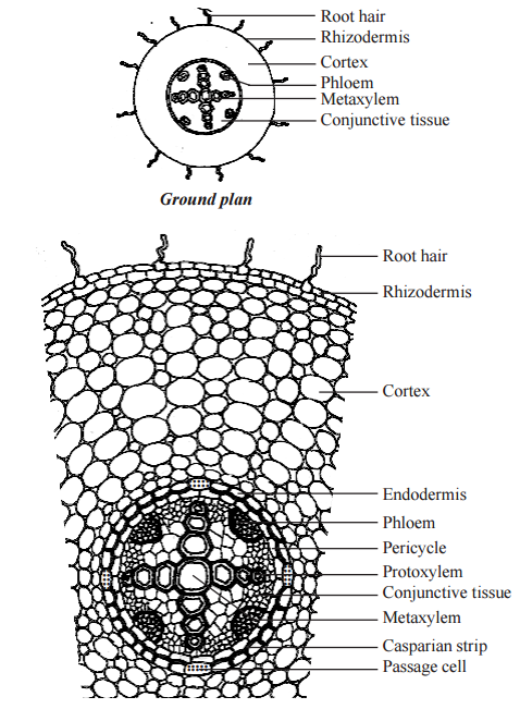
Cortex
Cortex consists of only parenchyma cells. These cells are loosely arranged with intercellular spaces to make gaseous exchange easier.
These cells may store food reserves. The cells are oval or rounded in shape.
Sometimes they are polygonal due to mutual pressure. Though chloroplasts are absent in the cortical cells, starch grains are stored in them.
The cells also possess leucoplasts.
The inner most layer of the cortex is endodermis. Endodermis is made up of single layer of barrel shaped parenchymatous cells.
Stele is completely surrounded by the endodermis. The radial and the inner tangential walls of endodermal cells are thickened with suberin.
This thickening was first noted by Casparay. So these thickenings are called Casparian strips.
But these casparian strips are absent in the endodermal cells which are located opposite to the protoxylem elements.
These thin-walled cells without casparian strips are called passage cells through which water and mineral salts are conducted from the cortex to the xylem elements.
Water cannot pass through other endodermal cells due to the presence of casparian thickenings.
Stele
All the tissues present inside endodermis comprise the stele. It includes pericycle and vascular system.
Pericycle
Pericycle is generally a single layer of parenchymatous cells found inner to the endodermis.
It is the outermost `layer of the stele. Lateral roots originate from the pericycle. Thus, the lateral roots are endogenous in origin.
Vascular system
Vascular tissues are in radial arrangement. The tissue by which xylem and phloem are separated is called conjunctive tissue.
In bean, the conjunctive tissue is composed of parenchymatous tissue. Xylem is in exarch condition.
The number of protoxylem points is four and so the xylem is called tetrarch. Each phloem patch consists of sieve tubes, companion cells and phloem parenchyma.
Metaxylem vessels are generally polygonal in shape. But in monocot roots they are circular.
For more details about dicotyledonous root click here
Describe the primary structure of a dicot root.
Draw the transverse section of dicot root and label the parts.
Explain the Primary structure of dicotyledonous root.
Other links
Plant anatomy – Meristematic tissue
Permanent tissue , simple tissue characteristics
Complex tissues , Xylem and its Kinds
Tissue system – Epidermal,Vascular and fundamental tissue system
Primary structure of monocotyledonous root – Maize root
Anatomy of monocot stem – Maize stem
Primary structure of dicotyledonous stem – Sunflower stem
Anatomy of a dicot and monocot leaves